
How Vitamin Deficiencies Accelerate Aging: Science Guide
Discover how vitamin deficiencies accelerate aging at the cellular level. Understand the key micronutrients affecting your health and longevity.
How Vitamin Deficiencies Accelerate Aging: Science Guide
Specific micronutrient deficiencies measurably accelerate biological aging through mitochondrial impairment, oxidative stress, impaired DNA repair, and epigenetic dysregulation. This is not a correlation story. The mechanisms are documented at the cellular level, and the nutrients most clearly implicated are vitamin D, B12, folate, vitamin C, vitamin E, magnesium, zinc, iron, and selenium. Micronutrient deficiencies affect a very large number of people globally and are directly linked to accelerated degenerative processes associated with aging.
Here is what the evidence shows at a glance:
- Vitamin D: Deficiency impairs autophagy, accelerates tissue aging, and is linked to frailty, cognitive decline, and immune dysfunction.
- B12 and folate: Shortfalls disrupt DNA methylation and nucleotide synthesis, pushing epigenetic age forward.
- Vitamin C and E: Antioxidant roles protect proteins, lipids, and DNA from reactive oxygen species (ROS); deficiency accelerates oxidative damage.
- Magnesium, zinc, iron, selenium: Cofactors for mitochondrial enzymes and antioxidant systems; deficits impair energy production and increase oxidant leak.
- Vitamin A: Deficiency harms immune function and tissue maintenance, but excess is linked to accelerated epigenetic aging.
- Evidence strength: Mitochondrial and oxidative mechanisms are well-established in lab studies; epigenetic links are emerging from large cohort analyses; randomized controlled trials (RCTs) measuring DNA methylation age are still limited.
Testing matters as much as knowing which nutrients to watch. A serum 25-hydroxyvitamin D, methylmalonic acid, homocysteine, ferritin, and serum zinc panel gives you objective data, not guesswork. The TIMELESS four-layer protocol pairs targeted nutrient correction with mitochondrial, emotional, spiritual, and energetic work because aging is a four-layer problem. Fixing one layer without the others leaves the program running.
Table of Contents
- How do vitamin deficiencies accelerate aging at the cellular level?
- Which specific nutrients speed up biological aging when they are low?
- What does the research actually prove about nutrients and aging?
- Who is most at risk for deficiencies that accelerate aging?
- How do you test for deficiencies and correct them safely?
- How quickly can you expect measurable changes after correcting deficiencies?
- Key Takeaways
- Why nutrient correction alone is not enough to reverse biological age
- What comes next: verified resources from Timeless - Reverse Your Age
- Useful sources and further reading
- FAQ
How do vitamin deficiencies accelerate aging at the cellular level?
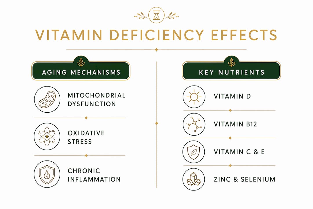
Nutrient shortfalls do not simply make you feel tired. They disrupt the molecular machinery that keeps cells functioning, replicating, and cleaning up damage. Five core mechanisms explain most of the biology.
Mitochondrial dysfunction
Mitochondria require a continuous supply of mineral and B-vitamin cofactors to run the electron transport chain efficiently. When iron or copper is insufficient, heme biosynthesis and Complex IV assembly are impaired, increasing mitochondrial oxidant leak and directly accelerating DNA damage. B vitamins (B1, B2, B3, B5, B6) serve as coenzymes in the citric acid cycle; without them, ATP production drops and reactive oxygen species accumulate faster than antioxidant systems can neutralize them. Magnesium is required for ATP stability itself. The result is a mitochondrial environment that ages cells faster than the calendar does. For a deeper look at how mitochondrial energy metabolism drives aging, the mechanisms go well beyond simple fatigue.
Oxidative stress and antioxidant defenses

Vitamins C and E, selenium (as glutathione peroxidase), and zinc (as superoxide dismutase) form the front line against ROS. Vitamin C regenerates oxidized vitamin E, so a deficiency in either one amplifies damage from the other. Selenium deficiency specifically reduces glutathione peroxidase activity, leaving lipid membranes and mitochondrial DNA exposed to peroxidation. The downstream effect is accelerated protein carbonylation, lipid peroxidation, and strand breaks in nuclear DNA.
Autophagy and proteostasis
Autophagy is the cell’s recycling system. It removes dysfunctional mitochondria (mitophagy) and protein aggregates before they accumulate into the debris associated with cellular senescence. Vitamin D regulates autophagy pathways that protect against cellular aging, and deficiency is linked to accelerated tissue aging in experimental and observational studies. Without adequate autophagy, senescent cells accumulate, secreting pro-inflammatory signals that damage neighboring tissue.
Chronic inflammation and inflammaging
Deficiencies in vitamin D, vitamin A, and zinc each impair immune regulation in ways that tip the balance toward chronic low-grade inflammation, a process researchers call “inflammaging.” This sustained inflammatory state drives the senescence-associated secretory phenotype (SASP), where damaged cells release cytokines that accelerate aging in surrounding tissue. Supporting immune system resilience through adequate micronutrient status is one of the most direct levers available.
Epigenetic and DNA-repair pathways
B12 and folate supply the one-carbon units needed for DNA methylation and nucleotide synthesis. When these nutrients are low, methylation patterns shift, gene expression changes, and epigenetic age accelerates. Folate deficiency also impairs thymidylate synthesis, causing uracil misincorporation into DNA and increased strand breaks. The Ames triage hypothesis proposes that when micronutrients are scarce, the body prioritizes short-term survival functions over long-term DNA repair and maintenance, meaning chronic low-level deficiency quietly accelerates aging even without overt clinical symptoms.
Key mechanism summary: Deficiency → impaired cofactor supply → mitochondrial oxidant leak + reduced antioxidant capacity + disrupted methylation + impaired autophagy → accelerated DNA damage, epigenetic age shift, and cellular senescence.
Which specific nutrients speed up biological aging when they are low?
The table below maps each nutrient to its primary aging mechanism, the lab tests used to detect deficiency, correction routes, and the strength of the evidence linking deficiency to accelerated aging.
| Nutrient | Primary aging mechanism | Key biomarkers/tests | Correction routes | Evidence strength |
|---|---|---|---|---|
| Vitamin D | Autophagy impairment, immune dysregulation, inflammaging | Serum 25(OH)D; sufficiency threshold ~30 ng/mL | Sun exposure, D3 supplementation, fatty fish | Strong observational; mechanistic lab data |
| Vitamin B12 | Epigenetic methylation disruption, neurodegeneration | Serum B12, methylmalonic acid (MMA), homocysteine | Oral high-dose B12, sublingual, or IM injection | Strong mechanistic; cohort data |
| Folate | DNA methylation, nucleotide synthesis, strand-break repair | Serum folate, red blood cell folate, homocysteine | Leafy greens, fortified foods, folic acid/methylfolate supplements | Strong mechanistic; epidemiological |
| Vitamin C | Antioxidant defense, collagen synthesis, ROS neutralization | Plasma ascorbate | Citrus, berries, bell peppers; supplementation | Mechanistic; limited RCT aging data |
| Vitamin E | Lipid peroxidation protection, membrane integrity | Plasma alpha-tocopherol | Nuts, seeds, olive oil; supplementation | Mechanistic; mixed RCT results |
| Magnesium | ATP stability, mitochondrial enzyme cofactor, DNA repair | Serum magnesium (note: poor reflection of total body stores); RBC magnesium more sensitive | Leafy greens, nuts, legumes; magnesium glycinate/malate | Mechanistic; epidemiological |
| Zinc | Superoxide dismutase activity, immune regulation, DNA repair | Serum zinc, alkaline phosphatase | Meat, shellfish, legumes; zinc picolinate/gluconate | Mechanistic; observational |
| Iron | Complex IV assembly, heme biosynthesis, oxygen transport | Ferritin, serum iron, transferrin saturation | Red meat, legumes; oral iron; IV iron when indicated | Strong mechanistic (Complex IV) |
| Selenium | Glutathione peroxidase activity, thyroid hormone activation | Serum selenium, plasma selenoprotein P | Brazil nuts (1–2/day), seafood; selenomethionine | Mechanistic; epidemiological |
| Vitamin A | Immune homeostasis, tissue maintenance | Serum retinol | Liver, dairy, eggs; beta-carotene from plants | Mechanistic; non-linear effects (see below) |
Vitamin A and B12: the non-linear warning. A 2025 analysis published in Frontiers in Nutrition found that higher serum vitamin A concentrations were associated with accelerated epigenetic aging, with an inflection point at approximately 71.5 μg/dL, while sufficient vitamin B12 (around 488.0 pg/mL) was linked to delayed epigenetic age acceleration. This is a critical finding: more is not always better. Blanket high-dose vitamin A supplementation without confirmed deficiency can push epigenetic age in the wrong direction.
A large NHANES analysis confirmed the multi-nutrient picture, finding that combined intake of protein, vitamin E, B vitamins, magnesium, iron, zinc, and other nutrients was negatively associated with accelerated phenotypic aging. No single nutrient explains the full picture. The aging program runs on a full stack of cofactors, and gaps anywhere in that stack create compounding damage.
Hormonal pathways matter too. Selenium deficiency impairs the conversion of T4 to active T3 thyroid hormone, slowing metabolic rate and amplifying mitochondrial inefficiency. Zinc and vitamin D both modulate testosterone and estrogen signaling. When these nutrients are low, the hormonal environment shifts toward one that accelerates tissue breakdown and reduces anabolic repair capacity.
What does the research actually prove about nutrients and aging?
The evidence base is strong in some areas and genuinely incomplete in others. Knowing the difference protects you from both under-correcting and over-supplementing.
What is well-established:
- Mechanistic lab studies clearly show that mineral deficits impair mitochondrial Complex IV, increase oxidant leak, and accelerate DNA damage. This is causal biology, not correlation.
- The Ames triage hypothesis provides a coherent framework: the body rations scarce micronutrients toward immediate survival, sacrificing long-term DNA repair. This predicts that chronic subclinical deficiency accelerates aging even without acute disease.
- Berridge (2017) proposed that vitamin D deficiency accelerates aging partly through impaired autophagy, supported by experimental evidence in animal models and cell culture.
- The NHANES phenotypic age acceleration data shows population-level associations between multi-nutrient intake patterns and biological aging outcomes.
- The Frontiers 2025 epigenetic analysis adds a non-linear dimension: vitamin A excess and B12 sufficiency have opposite effects on methylation-based aging clocks.
Where the evidence is weaker:
- Most human data is cross-sectional or cohort-based, meaning confounding is a real problem. People who are deficient in multiple nutrients often have other health challenges that independently accelerate aging.
- Bioavailability varies widely between individuals, making it hard to translate population-level intake data into individual dosing recommendations.
- RCTs that measure DNA methylation age as a primary outcome after correcting specific deficiencies are still rare. Most trials use surrogate endpoints like bone density or cognitive scores.
- Dose-response relationships are often U-shaped, not linear. The vitamin A finding is the clearest example, but similar non-linearity likely exists for iron and possibly selenium.
Open questions that would change practice:
- Large RCTs measuring TruAge or GrimAge clock changes after correcting individual deficiencies with controlled dosing.
- Studies separating the effect of correcting a deficiency from the effect of supplementing beyond sufficiency.
- Controlled trials examining whether improving absorption (rather than just increasing intake) produces better epigenetic outcomes.
- Data on whether the order of correction matters when multiple deficiencies coexist.
Who is most at risk for deficiencies that accelerate aging?
Knowing you should eat well is not the same as knowing whether your body is actually absorbing what you eat. Several physiological and pharmacological factors make deficiency likely even when intake looks adequate on paper.

Age-related absorption changes are the most underappreciated risk factor. Reduced gastric acid production, declining skin synthesis of vitamin D, and impaired intestinal and renal absorption make older adults especially prone to deficiencies in B12, vitamin D, and calcium even when dietary intake seems sufficient. Skin 7-dehydrocholesterol concentrations drop by more than 50% between ages 20 and 80, cutting vitamin D synthesis by roughly 40% in older adults. Reduced gastric acid also impairs the release of B12 from food-bound proteins, so serum B12 can look borderline while methylmalonic acid (a functional marker) is already elevated.
High-risk groups to know:
- Older adults (65+): Reduced acid, motility, skin synthesis, and renal conversion all converge. Deficiencies in vitamin D, B12, vitamin E, and selenium are common, with some studies showing inadequacies exceeding 80% for vitamin D, E, magnesium, and calcium in this group.
- People on proton pump inhibitors (PPIs) or H2 blockers: These drugs reduce gastric acid and directly impair B12 and magnesium absorption.
- Metformin users: Metformin interferes with B12 absorption in the ileum; long-term users should monitor B12 and MMA annually.
- Bariatric surgery patients: Bypass procedures reduce absorptive surface area for iron, B12, folate, and fat-soluble vitamins.
- Strict vegans and vegetarians: B12 is found almost exclusively in animal products. Zinc and iron from plant sources have lower bioavailability due to phytate binding.
- People with IBD or celiac disease: Chronic gut inflammation impairs absorption across multiple nutrient categories simultaneously.
- Individuals with chronic inflammation or multimorbidity: Elevated inflammatory cytokines suppress appetite (via cholecystokinin and leptin signaling) and impair nutrient utilization even when intake is technically adequate.
Symptoms that should trigger testing:
- Persistent fatigue not explained by sleep or workload
- Tingling, numbness, or balance issues (B12 neuropathy)
- Muscle weakness or bone pain (vitamin D)
- Slow wound healing or frequent infections (zinc, vitamin C)
- Hair thinning or brittle nails (iron, selenium, zinc)
- Cognitive fog or mood changes (B12, folate, vitamin D)
Pro Tip: When ordering labs for an aging workup, request serum 25(OH)D, serum B12 plus methylmalonic acid (MMA is the functional marker, not just serum B12), ferritin with transferrin saturation, serum zinc, serum selenium, and a homocysteine level. This panel catches functional deficiencies that standard CBC and metabolic panels miss entirely.
How do you test for deficiencies and correct them safely?
Testing before supplementing is not optional. The vitamin A data alone makes that clear: supplementing without confirmed deficiency can accelerate epigenetic aging rather than slow it.
Baseline testing roadmap for an aging workup:
- Serum 25(OH)D: Target sufficiency at 30 ng/mL or above; many clinicians in aging medicine aim for sufficiency thresholds commonly used in practice.
- Serum B12 + methylmalonic acid: Serum B12 alone misses functional deficiency. MMA rises before neurological symptoms appear.
- Homocysteine: Elevated homocysteine signals functional B12 or folate insufficiency and is an independent cardiovascular and cognitive risk marker.
- Ferritin + transferrin saturation: Ferritin below 30 ng/mL suggests depleted stores even when hemoglobin is normal.
- Serum zinc: Fasting morning draw is most reliable; values below 70 μg/dL warrant attention.
- Serum selenium: Plasma selenoprotein P is the most sensitive marker but less commonly ordered; serum selenium is a reasonable proxy.
- RBC magnesium: More sensitive than serum magnesium for detecting intracellular depletion.
Correction routes and safety notes:
- Vitamin D: Oral D3 with vitamin K2 (to direct calcium appropriately) is the standard correction route. Recheck 25(OH)D at 8–12 weeks after starting supplementation.
- B12: Oral high-dose cyanocobalamin or methylcobalamin works for most people. Intramuscular injection is reserved for confirmed malabsorption (pernicious anemia, post-bariatric). Recheck serum B12 and MMA at 3 months.
- Iron: Oral ferrous bisglycinate is better tolerated than ferrous sulfate. IV iron (ferric carboxymaltose) is appropriate when oral iron fails or is contraindicated. Recheck ferritin at 8–12 weeks.
- Folate: Methylfolate (5-MTHF) is preferred over folic acid for individuals with MTHFR variants. Dietary sources include dark leafy greens, lentils, and asparagus.
- Magnesium: Glycinate and malate forms have better absorption and GI tolerance than oxide. Take separately from calcium supplements.
- Zinc: Separate from iron supplements by at least 2 hours; they compete for the same transporter. Zinc picolinate and gluconate are well-absorbed forms.
Absorption optimization matters as much as dose. Aging-related digestive changes mean clinicians must treat absorption, not just intake. Betaine HCl support for low stomach acid can meaningfully improve B12 and mineral absorption in older adults. Fat-soluble vitamins (D, A, E, K) absorb best with a fat-containing meal. Vitamin C taken with non-heme iron sources roughly doubles iron absorption.
Safety cautions:
- Never supplement vitamin A at high doses without confirmed deficiency and clinician oversight. The epigenetic aging data is unambiguous on this point.
- High-dose iron supplementation without confirmed deficiency increases oxidative stress and is contraindicated.
- High-dose zinc (above 40 mg/day long-term) can deplete copper; monitor copper status if zinc repletion is prolonged.
- Fat-soluble vitamins accumulate; toxicity is possible with sustained excess doses of D, A, and E.
Pro Tip: Use functional follow-up tests rather than symptom monitoring alone. Repeat MMA and homocysteine at 3 months after starting B12 and folate correction. Repeat 25(OH)D at 8–12 weeks. Ferritin at 8–12 weeks. These functional markers tell you whether the correction is actually working at the cellular level, not just whether the serum level moved.
How quickly can you expect measurable changes after correcting deficiencies?
Correction timelines vary by nutrient, severity of deficiency, and the individual’s absorption capacity. Here is what the evidence supports:
Short-term (days to 2 weeks):
- Energy levels and mood often improve within days of correcting severe B12 or iron deficiency.
- Neurological symptoms from B12 deficiency (tingling, balance issues) may begin to stabilize within 1–2 weeks of injection therapy, though full recovery takes longer.
- Vitamin C repletion can improve wound healing markers within days in severely depleted individuals.
Medium-term (4–12 weeks):
- Serum 25(OH)D reaches a new steady state at 8–12 weeks after a consistent D3 dose. Retest at this point.
- Ferritin recovers to normal range in 8–12 weeks with consistent oral iron in most adults without ongoing blood loss.
- Homocysteine levels typically fall within 4–8 weeks of adequate B12 and folate repletion, reflecting improved methylation capacity.
- Skin quality changes linked to vitamin C and zinc correction (collagen synthesis, reduced oxidative damage) become noticeable at 8–12 weeks.
Longer-term (3–12 months):
- Epigenetic age markers (GrimAge, PhenoAge, TruAge) require at least 3–6 months of sustained correction to show meaningful shifts. Single-nutrient corrections in isolation produce modest changes; multi-layer interventions produce larger ones.
- Muscle strength and bone density markers respond over 6–12 months of sustained vitamin D and protein sufficiency.
- Cognitive screening scores in B12-deficient older adults can improve measurably over 3–6 months of repletion, though this depends heavily on how long the deficiency was present before correction.
Clinical endpoints worth tracking alongside labs:
- Grip strength (a validated proxy for biological age and sarcopenia risk)
- Gait speed and balance tests
- Cognitive screening (MoCA or similar)
- Bone turnover markers (CTX, P1NP) for vitamin D response
- DNA methylation testing at 6-month intervals to objectively verify whether biological age is moving
Individual variability is real. Two people with the same serum B12 level can have very different functional outcomes depending on their MTHFR status, gut microbiome, and inflammatory load. This is why objective biomarker tracking beats symptom-only monitoring every time.
Key Takeaways
Specific micronutrient deficiencies accelerate biological aging through mitochondrial impairment, oxidative stress, epigenetic disruption, and chronic inflammation, and correcting them with targeted, tested protocols produces measurable changes in aging biomarkers within weeks to months.
| Point | Details |
|---|---|
| Priority nutrients | Vitamin D, B12, folate, vitamin C, E, magnesium, zinc, iron, and selenium each drive distinct aging mechanisms. |
| Non-linear risk | Higher serum vitamin A is linked to accelerated epigenetic aging; supplement only confirmed deficiencies, not preemptively. |
| Test functionally | Serum B12 alone misses deficiency; add methylmalonic acid and homocysteine for a true picture of cellular status. |
| Correction timelines | Biomarkers shift in 4–12 weeks; epigenetic age markers require 3–6 months of sustained multi-layer correction. |
| Timeless - Reverse Your Age | The four-layer TIMELESS protocol pairs nutrient correction with mitochondrial, emotional, spiritual, and energetic work, verified by TruAge DNA methylation testing. |
Why nutrient correction alone is not enough to reverse biological age
Most longevity protocols stop at the physical layer. Fix the vitamin D, optimize the B12, eat more leafy greens. That is necessary work, and the research reviewed here makes clear it is not optional. But in my clinical experience working with thousands of practitioners across 40 countries, I have seen people with near-perfect lab panels who still test biologically older than their chronological age.
The reason is that aging is a four-layer problem. The Physical layer covers cellular, epigenetic, and mitochondrial function. That is where nutrient correction lives. But the Emotional layer, stored trauma and chronic nervous system dysregulation, keeps cortisol and inflammatory signaling elevated in ways that no supplement can override. The Spiritual layer, the identity-level beliefs you hold about your own aging, shapes the epigenetic expression of those beliefs through well-documented psychoneuroimmunological pathways. The Energetic layer, the bioelectric field and meridian system, modulates cellular communication in ways that are measurable but rarely addressed in conventional protocols.
The TIMELESS protocol addresses all four layers simultaneously. My own TruAge DNA methylation test confirmed a biological age of 23 at chronological age 41. That result did not come from optimizing one layer. It came from treating aging as the multi-dimensional program it actually is, and rewriting each layer with verified, evidence-backed methods. Correcting your deficiencies is the right first move. Pairing that correction with the full four-layer approach is what produces results that show up on a DNA methylation clock.
What comes next: verified resources from Timeless - Reverse Your Age
If the mechanisms covered here have you ready to act, there are three concrete next steps available right now.
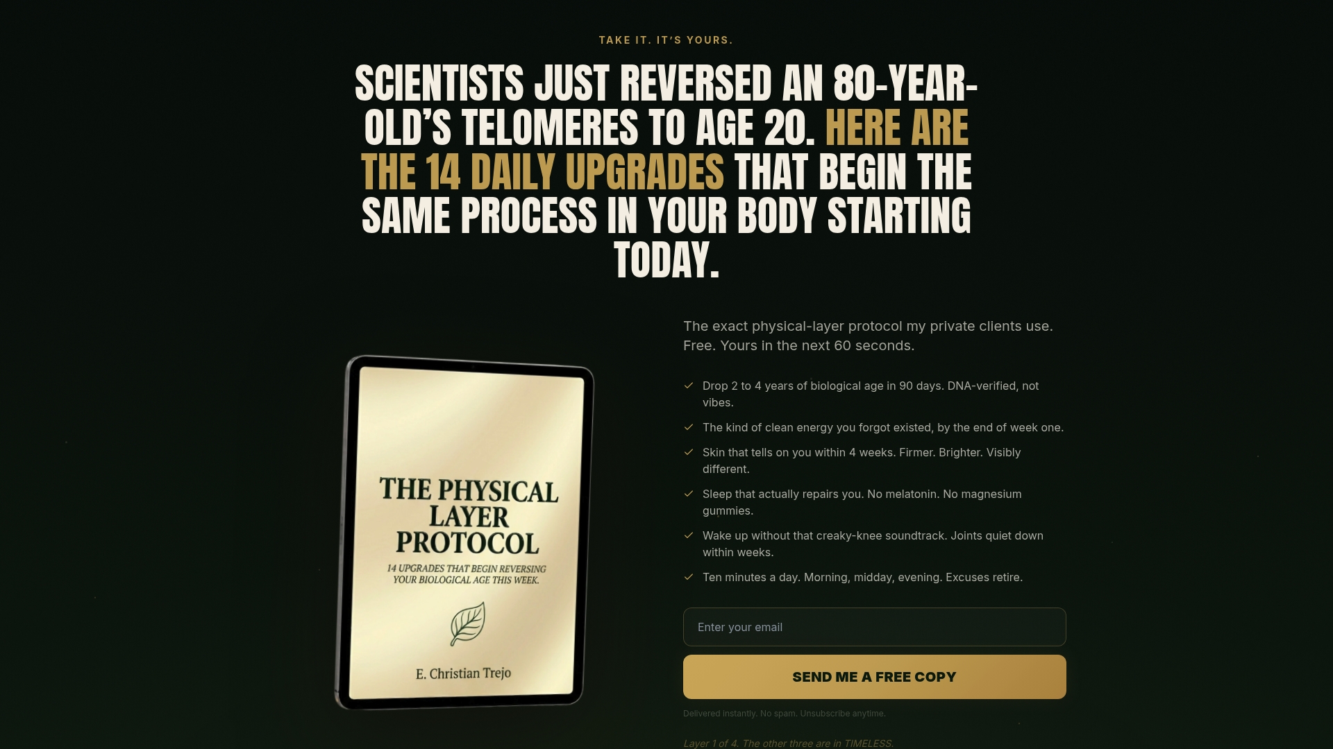
The free guide at reverseyourage.org/14-upgrades covers 14 specific protocol upgrades that address all four aging layers, including the nutrient correction strategies outlined in this article. It costs nothing and gives you a working framework immediately.
The TIMELESS book ($29.99, paperback) is the 370-page blueprint documenting the full four-layer protocol, built on 15 years of research and tested across 40 countries. Every mechanism discussed in this article is expanded with protocols, dosing frameworks, and the science behind each intervention.
For readers who want a guided, DNA-verified program, the TIMELESS Vitality Intensive is an 8-week private coaching program that includes TruAge DNA methylation testing at entry and exit. The guarantee is straightforward: reverse biological age by at least 10 years in 6 months, verified by DNA testing, or every dollar is refunded. No other longevity program offers that level of accountability. Book a free Vitality Diagnosis call at reverseyourage.org/book to find out whether you qualify.
Useful sources and further reading
The following peer-reviewed studies and reviews form the primary evidence base for this article. All are accessible via PubMed or PMC.
-
Mechanistic insights into mitochondrial dysfunction from mineral deficiency (PubMed 16102804): Documents how iron and copper shortfalls impair Complex IV assembly, increase mitochondrial oxidant leak, and accelerate DNA damage. The foundational mechanistic citation for the mitochondrial section.
-
Non-linear association between serum vitamins A and B12 and accelerated epigenetic aging (Frontiers in Nutrition, 2025): Identifies inflection points for vitamin A (~71.5 μg/dL) and B12 (~488.0 pg/mL) in relation to PhenoAge and GrimAge acceleration. Critical for understanding why blanket supplementation is risky.
-
The association between dietary nutrient intake and acceleration of aging: evidence from NHANES: Large population-level analysis showing combined intake of multiple nutrients is negatively associated with phenotypic age acceleration. Supports the multi-nutrient framing.
-
Main nutritional deficiencies: Documents the global scale of micronutrient deficiency and its links to mitochondrial decay and degenerative aging processes.
-
Nutritional and lifestyle management of the aging journey: A narrative review: Covers age-related absorption changes, the role of gastric acid decline in B12 and mineral deficiency, and the principle that clinicians must treat absorption, not just intake.
-
Micronutrient status and protein-energy malnutrition in free-living older adults: Comprehensive review of deficiency prevalence and health consequences in community-dwelling older adults, including the 50% decline in skin 7-dehydrocholesterol between ages 20 and 80.
-
Vitamin D deficiency accelerates aging and age-related diseases: Experimental and observational evidence linking vitamin D deficiency to accelerated tissue aging, supporting the autophagy and inflammaging mechanisms described in this article.
FAQ
What vitamin deficiency is most directly linked to faster biological aging?
Vitamin D and B12 deficiencies have the strongest combined evidence for accelerating biological aging. Vitamin D deficiency impairs autophagy and drives chronic inflammation, while low B12 disrupts DNA methylation patterns that directly shift epigenetic age forward.
Does correcting B12 levels make you look or feel younger?
Correcting a genuine B12 deficiency can improve skin quality, energy, cognitive clarity, and neurological function within weeks to months, because B12 is required for cell turnover, myelin maintenance, and methylation-dependent gene regulation. The effect depends on how depleted you were and how long the deficiency was present.
What is the best vitamin to slow biological aging?
No single vitamin does the job alone. The strongest evidence points to vitamin D (autophagy, immune regulation), B12 and folate (epigenetic methylation), and the antioxidant nutrients vitamin C, E, and selenium (oxidative protection). A multi-nutrient approach supported by lab testing outperforms any single supplement.
What vitamin helps tighten and repair aging skin?
Vitamin C is the most directly supported nutrient for skin structure, as it is required for collagen synthesis and protects against UV-induced oxidative damage. Zinc supports wound healing and cell turnover, while vitamin E protects membrane lipids from peroxidation. Deficiency in any of these accelerates visible skin aging.
Can you reverse biological aging by fixing nutrient deficiencies?
Correcting deficiencies is necessary but not sufficient on its own. Epigenetic age markers can shift meaningfully over 3–6 months of targeted correction, but the largest and most verified outcomes come from protocols that address all four layers of aging: physical, emotional, spiritual, and energetic simultaneously.